February 12, 2015. HOLTZ retinal and optic-disc analysis research generated new knowledge for the R&D team, supporting increasingly precise hardware and software for quantitative analysis of conditions such as glaucoma. The current phase investigated focus stacking to improve sharpness across different image regions. Danilo Motta, an ICMC-USP intern, was one of the project leaders.
Abstract control ID: 2149169. Title: Development of a Focus Stacking Algorithm for Improvement of 3D Retinal Image Reconstruction. Authors: Luis A. Carvalho, Danilo Motta and Valeria Carvalho. Institutional affiliation at submission: the R&D department and Universidade de São Paulo, São Carlos, São Paulo, Brazil. Commercial disclosures reported a personal financial interest for Luis Carvalho, a consulting relationship for Danilo Motta and employment for Valeria Carvalho with the companies involved at the time.
Purpose: Ophthalmology needs more cost-effective instrumentation for early glaucoma diagnosis. A previous low-cost 3D retinal camera showed promising results. However, accurate stereo-image cross-correlation is a major challenge in 3D reconstruction, and depth of field also plays an important role.
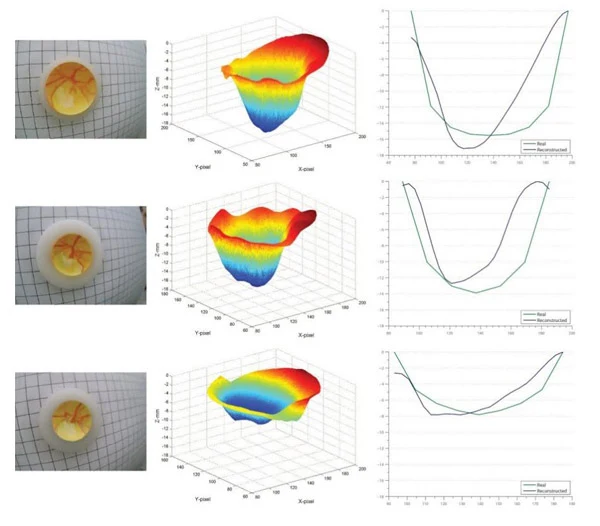
Methods: Focus stacking was applied to images acquired with the previously developed 3D retinal camera. Stereo videos of a model eye were acquired at progressive focusing planes using a CCD and zoom lens. A MATLAB algorithm preprocessed the video and selected image pairs with distinct focus characteristics. Edge detection and Fourier analysis identified in-focus regions automatically, and the sharp patches were blended into a final image.
Results: The algorithm was tested on model eyes with three optic-disc shapes manufactured to 5-micron precision. Results from the prior algorithm were compared before and after stacking. Root-mean-square error was 0.14 mm without stacking and 0.10 mm with stacking, an improvement of almost 30%.
Conclusions: In this preliminary model-eye study, focus stacking, originally used in photography and microscopy, improved 3D optic-disc reconstruction precision. The next goal was to apply the algorithms to in vivo eyes and compare them with more sophisticated and expensive optic-nerve depth-analysis techniques such as scanning laser ophthalmoscopy.
Reference: Carvalho LAV, Ramos E, Romano A. Construction of a True-depth Quantitative and Low Cost 3D Optical Disc Measurement Device and Collaborative Diagnosis using Stereo Images and Videos. ARVO, Investigative Ophthalmology & Visual Science 2012;53:E-Abstract 3102.
Lay summary: Many sophisticated glaucoma instruments are expensive and difficult to access in developing countries. The project aimed to develop simple, effective software that improves the precision of an affordable instrument intended to approach the performance of more expensive systems.
Submission details: Presentation preferences were poster, then paper. Reviewing code: 2330, imaging: new technologies and techniques — MOI. Section: Multidisciplinary Ophthalmic Imaging Group. Keywords: image processing (549), optic disc (627), retina (688). No clinical-trial registration was reported. Grant support was declared from the company’s US operation at the time.