THE PLATFORM’S DEVELOPMENT RECORD
Technology with a documented foundation.
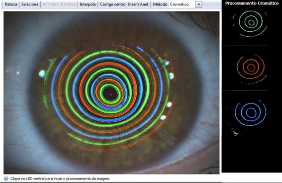
CHROMA combined videokeratography with anterior-corneal aberrometry. The team developed an image-processing approach that used chromatic Placido patterns as well as conventional monochromatic processing to reconstruct corneal shape and calculate optical aberrations.
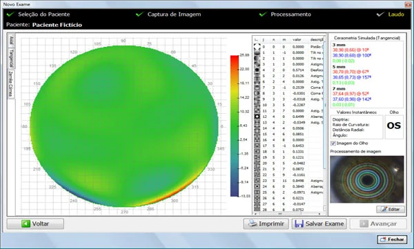
Earlier generations of topographers mainly calculated axial or instantaneous curvature and dioptric-power maps, with elevation calculated using methods such as Arc Step. Refraction maps were also available, but offered limited additional information beyond curvature-based power maps. Wider use of wavefront concepts and anterior-corneal wavefront algorithms opened a path to more detailed optical analysis. CHROMA applied these concepts in a corneal analysis system developed in Brazil.
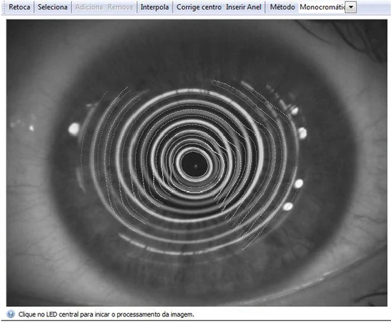
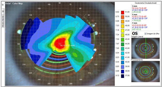
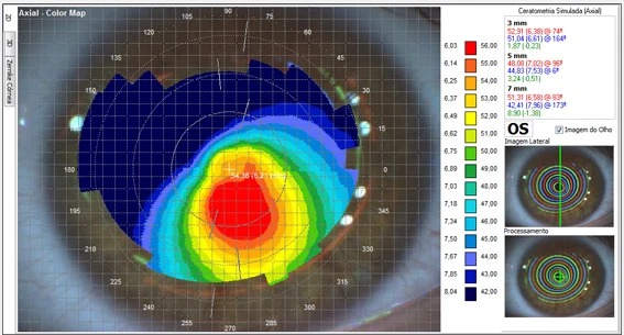
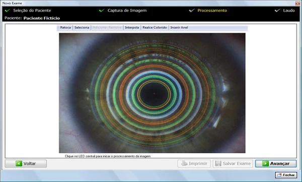
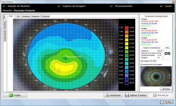
The software supported routine examinations, research, teaching, image processing and export for presentations. The archived comparison showed a keratoconic eye in which conventional monochromatic processing failed to detect portions of the Placido-ring boundaries, while the team's patented chromatic processing detected the previously missing regions. The accompanying images compare monochromatic and chromatic processing and their axial maps.
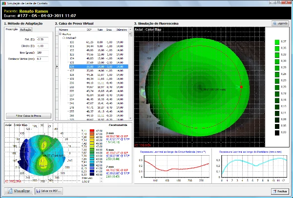
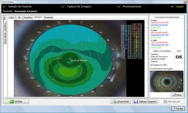
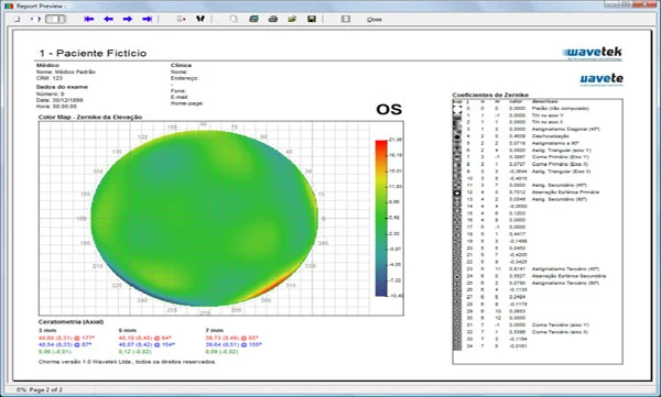
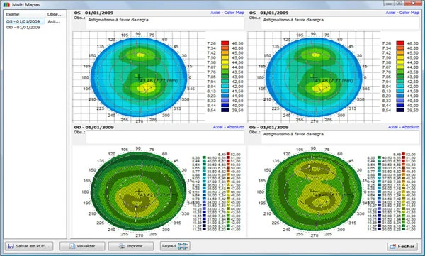
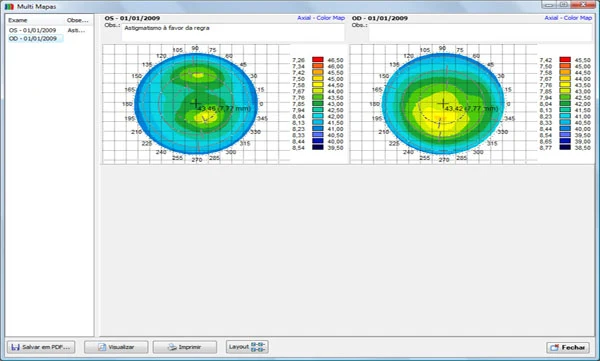
Software modules included axial curvature, tangential or instantaneous curvature, true elevation, Zernike-polynomial elevation fitting, Zernike-based anterior-corneal optical aberrations, contact-lens fitting with simulated fluorescein patterns, orthokeratology lens fitting, statistical analysis of corneal parameters, keratoconus statistics, multiple-map views and differential maps.
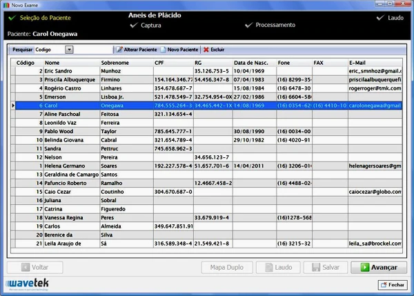
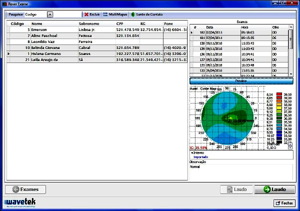
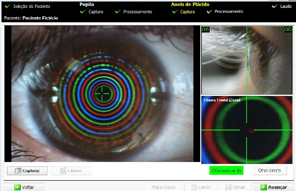
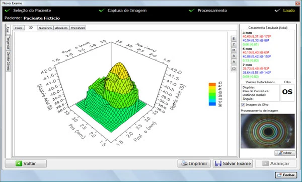
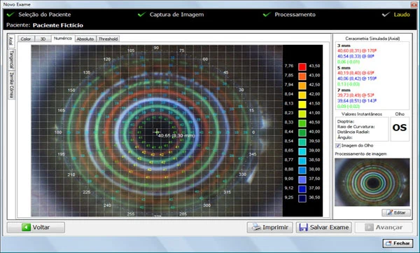
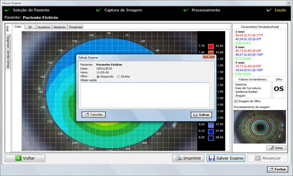
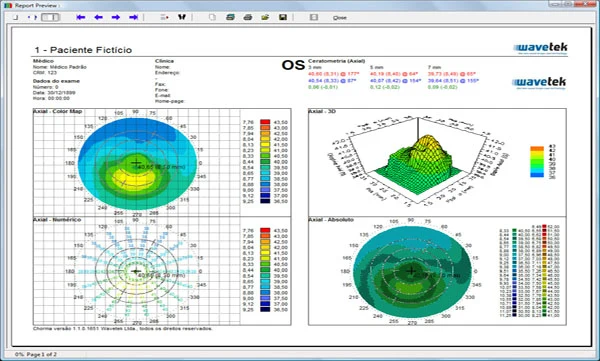
The patient and examination workflow included patient selection with examination previews, image capture, image processing, keratometry tables and two-dimensional plots, three-dimensional corneal maps and numerical reports showing dioptric power at selected points. The database stored the original Placido images as well as examination data.
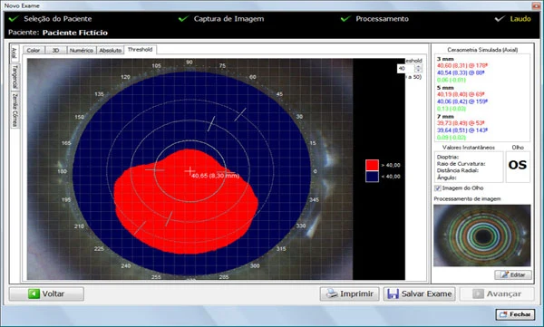
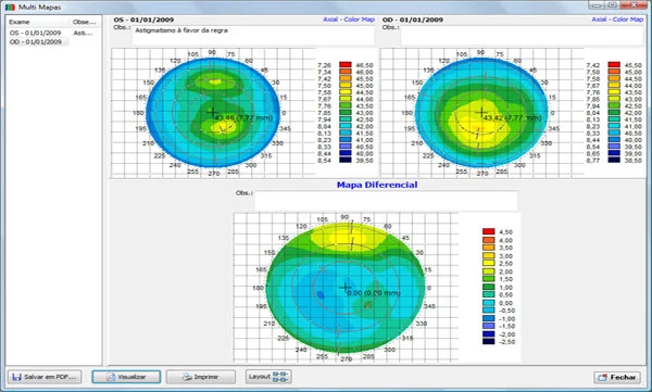
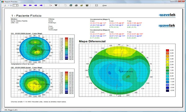
A threshold map let the clinician select a dioptric threshold and identify regions with power below that value in blue and above it in red. The software also displayed Zernike-fitted elevation maps and allowed simultaneous comparison of four examinations or two examinations from the same patient. Differential maps subtracted one examination from another.
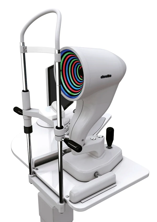
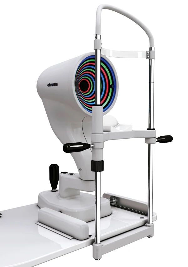
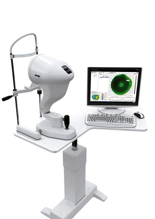
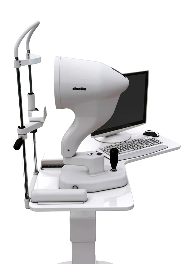
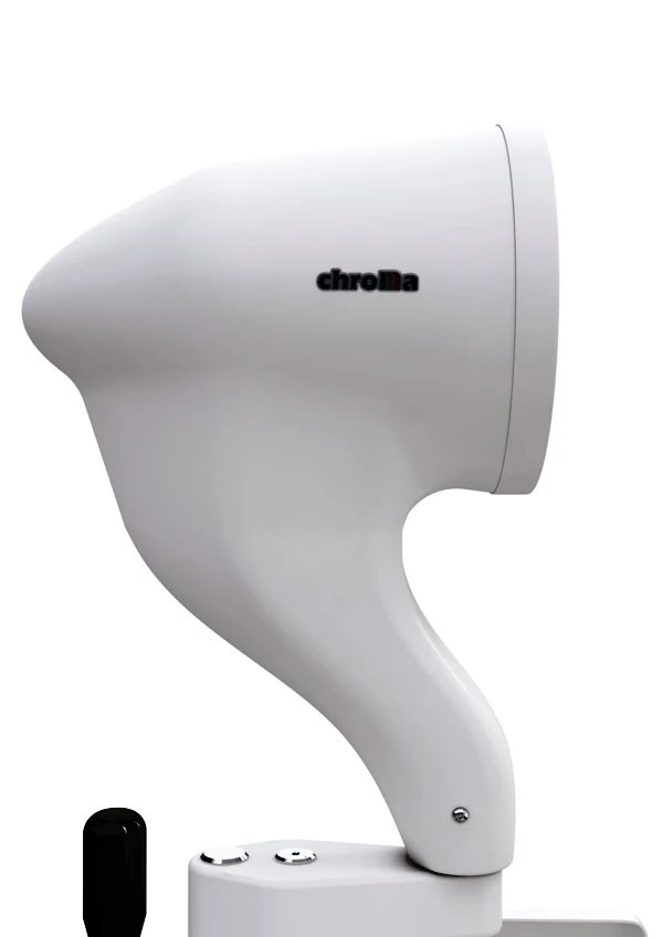
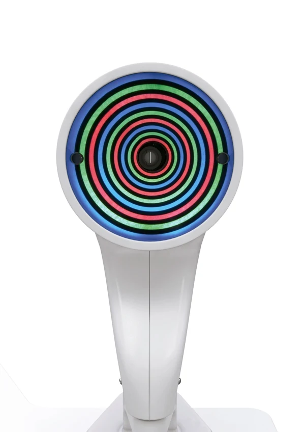
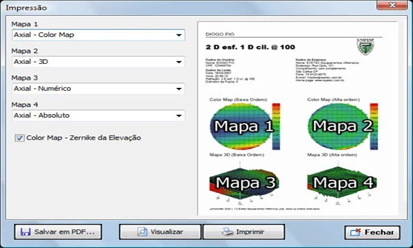
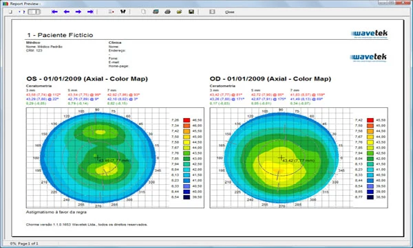
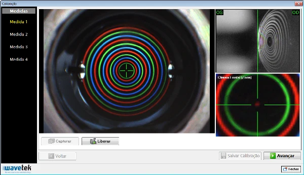
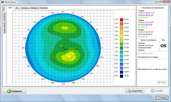
Report layouts and views could be configured before printing, with PDF output supported. Other archived examples show saved-examination review, examination storage and an instrument-calibration assistant. Product views include front, rear and side perspectives, and the source includes an automatic image-capture demonstration and a technical-specification PDF.
The original archive lists Brazilian ANVISA registration 80497810008 and makes historical statements about US FDA status. Those statements are retained only as context for the archived instrument, not as a claim that EVOptica holds a current authorization or certification.
Instrument, imaging & software gallery

BROCHURES / VIDEOS / SCIENTIFIC REFERENCES
Explore CHROMA.
English user manuals & guides
Technical-review editions of the full original instructions. Source differences and legacy interface labels are identified inside each document.
Instrument videos & recorded coverage
CHROMA automatic image capture demonstration
Watch the original video ↗Product recording / YOUTUBEValéria M. Carvalho on chromatic corneal topography and diagnostic innovation
Watch the original video ↗Related scientific publications
Foundational research related to this field. These articles are not clinical validation of a future instrument configuration.
Keratoconus prediction using a finite element model of the cornea with local biomechanical properties.
CARVALHO, LA ; PRADO, M. ; CUNHA, R. H. ; COSTA, A. ; PARANHOS JR, A. ; Schor ; Chamon .
Read the journal / DOI source ↗[Preliminary results of a wide-angle corneal topography system for Placido-based videokeratographers].
CARVALHO, LA .
Read the journal / DOI source ↗Neural networks and statistical analysis for classification of corneal videokeratography maps based on Zernike coefficients: a quantitative comparison
CARVALHO, LA ; BARBOSA, M.
Read the journal / DOI source ↗Mumford–Shah algorithm applied to videokeratography image processing and consequences to refractive power values
FLORINDO, J. B. ; SOARES, S. H. M. ; CARVALHO, LA ; Bruno . MUMFORD-SHAH ALGORITHM APPLIED TO VIDEOKERATOGRAPHY IMAGE PROCESSING AND CONSEQUENCES TO REFRACTIVE POWER VALUES.
Read the journal / DOI source ↗Desenvolvimento e resultados preliminares de um sistema para topografia da córnea utilizando iluminação através de fenda retangular estreita
CARVALHO, LA .
Read the journal / DOI source ↗The Placido Wavefront Sensor and Preliminary Measurement on a Mechanical Eye
CARVALHO, LA .
Read the journal / DOI source ↗A precise computer simulation system for rigid contact lens (PMMA) adaptation using in vivo data from corneal topography
CARVALHO, LA .
Read the journal / DOI source ↗[Techniques for facilitating in vivo corneal topography diagnosis].
CARVALHO, LA .
Read the journal / DOI source ↗Preliminary Results of Neural Networks and Zernike Polynomials for Classification of Videokeratography Maps
CARVALHO, LA .
Read the journal / DOI source ↗Spatial and frequency domain techniques for segmentation of Placido images and accuracy implications for videokeratography
CARVALHO, LA .
Read the journal / DOI source ↗Absolute accuracy of Placido-based videokeratographs to measure the optical aberrations of the cornea.
CARVALHO, LA .
Read the journal / DOI source ↗On the absolute accuracy of Zernike polynomials to characterize the corneal surface and the optical aberrations of the human eye
CARVALHO, LA .
Read the journal / DOI source ↗Videokeratoscopes for dioptric power measurement during surgery.
CARVALHO, LA ; STEFANI, M
Read the journal / DOI source ↗Videokeratograph (VKS) for monitoring corneal curvature during surgery
CARVALHO, LA ; Romão ; Tonissi ; Yasuoka ; Castro ; Schor ; Chamon .
Read the journal / DOI source ↗Desenvolvimento de um instrumento computadorizado para medida do poder refrativo da córnea (videoceratógrafo)
CARVALHO, LA .
Read the journal / DOI source ↗Computer Algorithm for Simulation of the Human Optical System and Contribution of the Cornea to the Optical Imperfections of the Eye
CARVALHO, LA .
Read the journal / DOI source ↗Development of a computerized instrument for evaluating human corneal curvature
CARVALHO, LA ; PARANAGUA, E. ; SANTOS, L. E. R. ; Tonissi ; Romão ; Oliveira ; Yasuoka ; Castro .
Read the journal / DOI source ↗Development of a computerized instrument for evaluating human corneal curvature
CARVALHO, LA ; SOUZA, S. J. F. E. ; Castro .
Read the journal / DOI source ↗Original instrument documents & sources
Brochures and manuals link to English EVOptica editions. Historical recordings, source citations and legacy interface screenshots may retain their original language. External archive links may no longer be available.