THE PLATFORM’S DEVELOPMENT RECORD
Technology with a documented foundation.
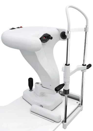
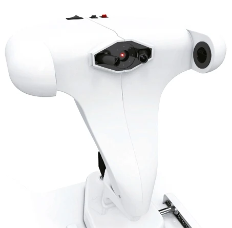
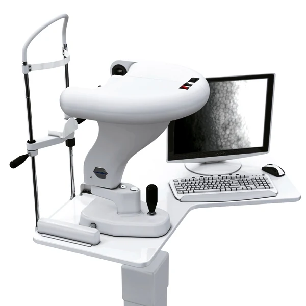
HERON brought together optical, mechanical, electronic and software development in a non-contact specular microscope for imaging and analyzing the corneal endothelium. The team developed the technology in Brazil, using electronic components sourced from Japan and optical components produced partly in São Carlos and partly in the United States.
The instrument was designed for routine ophthalmic examinations as well as research, teaching, image processing and export of images for presentations or online use. Its integrated computer architecture allowed software and computer hardware upgrades instead of requiring replacement of the complete instrument. The archived configuration used Windows XP or Windows Vista; these are historical platform specifications.
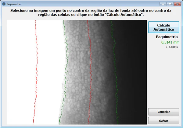
The optical pachymetry module calculated corneal thickness from the captured images. Thickness appeared in the existing reports and in a dedicated pachymetry report. The archived description reported agreement of approximately ±10% relative to ultrasound pachymetry.
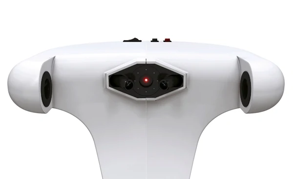
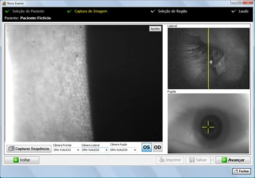
Image capture combined three alignment cameras—two lateral cameras and a central camera—with a fourth camera for endothelial imaging. Capture was controlled with a USB foot pedal. The operator could choose from eight pre-captured images or select suitable images from a sequence of up to 60 frames. The semi-automatic workflow let the clinician select the location and number of images to capture.
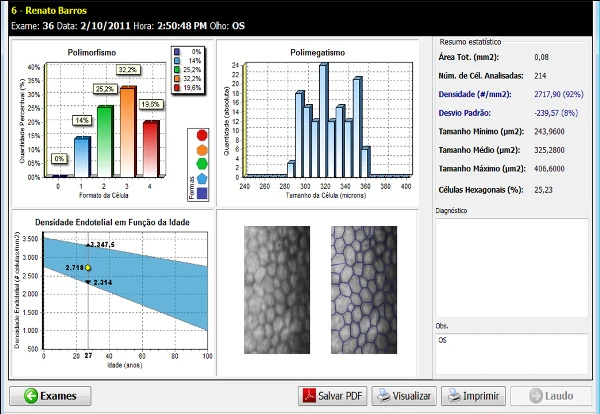
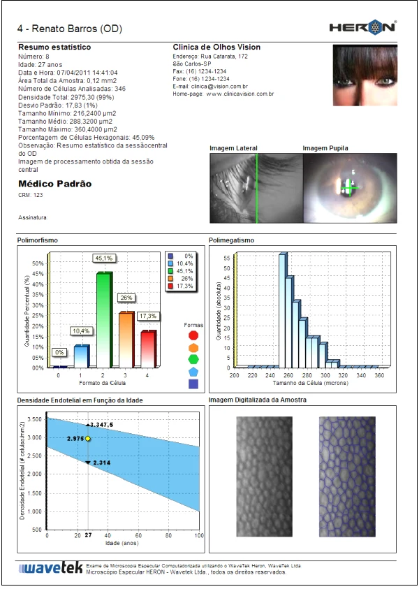
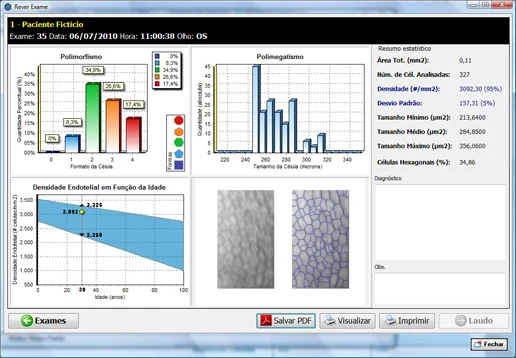
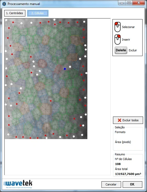
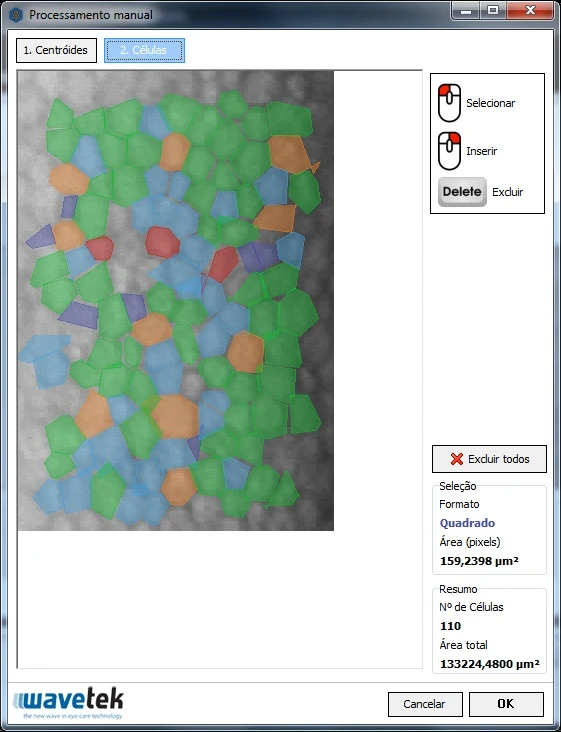
Automatic image processing detected cell boundaries, counted cells and calculated cell-size variation and cell-shape variation. Manual processing allowed the operator to make the measurements directly. The color display showed the cell images in real time during capture.
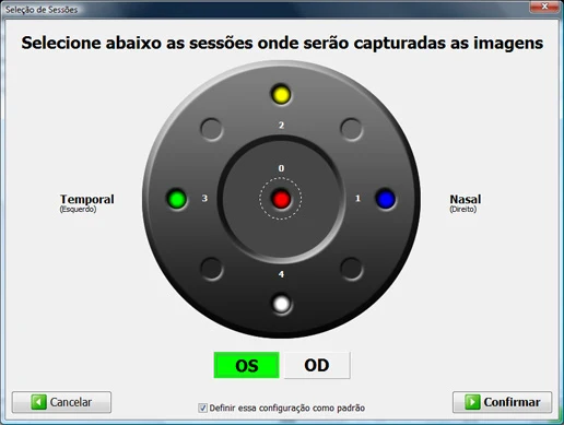
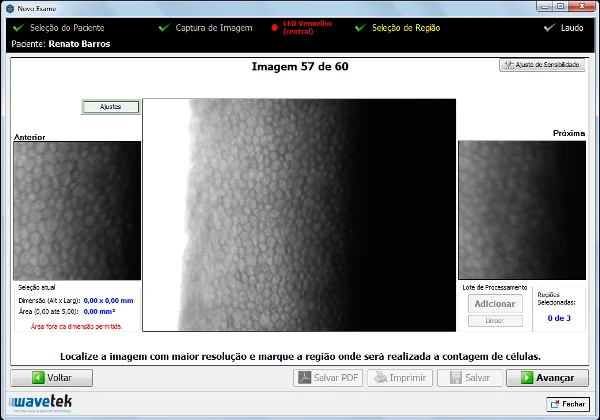
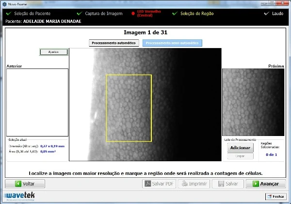
An LED fixation system supported five examination positions: central, temporal, nasal, superior and inferior. This multi-region approach allowed sampling from different areas of the corneal endothelium. Up to three regions of interest could be selected within an image for processing.
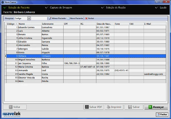
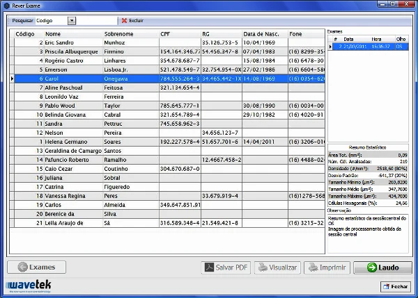
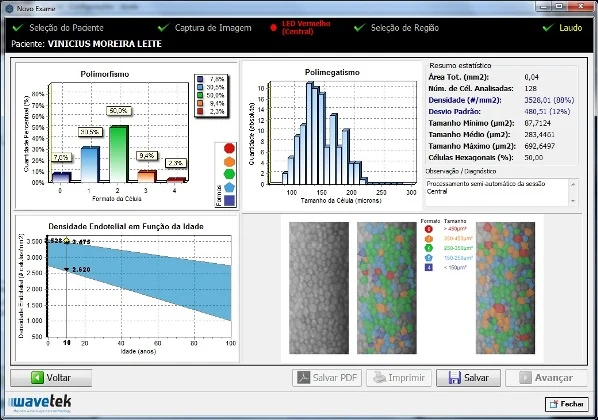
The patient and examination database stored images, cell measurements and patient, clinician and clinic details. Reports included cell density in cells/mm², cell-size and cell-shape statistics, and plots comparing results with a population of the same age group. Color A4 reports could include the lateral view and pupil image and could be printed or exported as PDF. Stored examinations could be reviewed, reprinted and exported. A one-click backup function supported data preservation.
Additional archived features included capture at different corneal locations, selection of regions and subregions for analysis, individual editing of cells, the number of cell sides and neighboring cells, color printing through compatible Windows printers, and a network viewer for reviewing and editing examinations. The four high-resolution color CCD cameras supported alignment and image acquisition. The instrument housing was developed with designers trained at UNESP and USP, and the software interface incorporated feedback from Brazilian ophthalmologists.
The historical comparison table also described slit-lamp coupling, remote technical support, software upgrades, population-based statistical comparisons, a training course with Dr. Fernando Abib and Brazilian development of the optics, electronics, mechanics and software. These describe the historical project and its support model rather than a current EVOptica product offer.
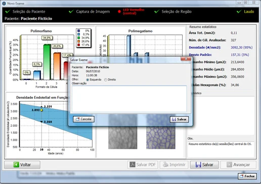
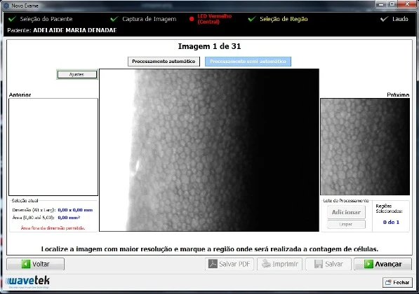
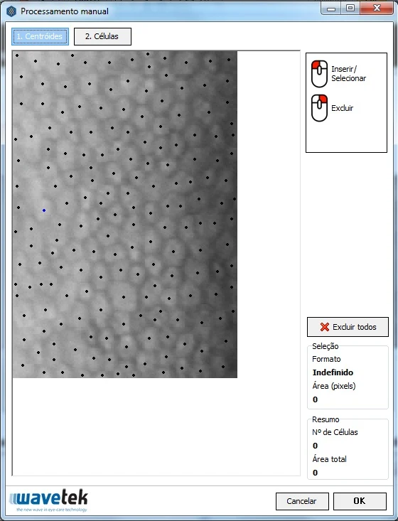
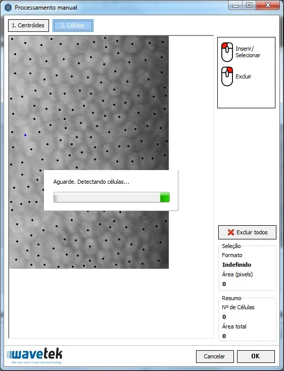
Manual processing workflow: first select the sharpest captured image. Mark the region for cell counting, choose Add and continue. In the manual processing screen, edit or delete the detected centroids or insert additional centroids. After defining the centroids, select the Cells step to detect cell boundaries. Edit, delete or add cells as needed, then confirm with OK to display the final report. This workflow let clinicians apply their experience when selecting cells for density, size-variation and shape-variation calculations.
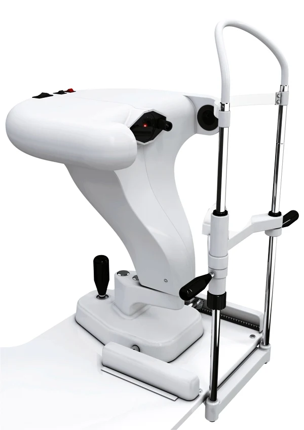
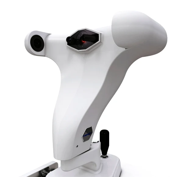
The source includes a technical-specification PDF, instrument views, capture examples, software screenshots and a historical comparison presentation. Regulatory and commercial statements in these original materials refer to the instrument and organization at the time; they do not establish current EVOptica certification or availability.
Instrument, imaging & software gallery

BROCHURES / VIDEOS / SCIENTIFIC REFERENCES
Explore HERON.
English user manuals & guides
Technical-review editions of the full original instructions. Source differences and legacy interface labels are identified inside each document.
Instrument videos & recorded coverage
HERON endothelial-cell image capture
Watch the original video ↗Product recording / YOUTUBEHERON manual endothelial-cell processing and individual cell editing
Watch the original video ↗Original instrument documents & sources
Brochures and manuals link to English EVOptica editions. Historical recordings, source citations and legacy interface screenshots may retain their original language. External archive links may no longer be available.