THE PLATFORM’S DEVELOPMENT RECORD
Technology with a documented foundation.
HOLTZ combined high-resolution retinal imaging with quantitative optic-disc analysis. The archive describes development of an optic-cup depth calculation and 3D visualization module in collaboration with a US company and the computer-science department at the University of California, Berkeley, where one of the founders had studied and maintained research connections. The accompanying video demonstrates the module.
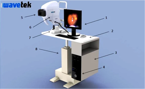
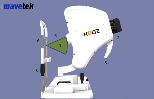
The system configuration included an LCD monitor, tabletop, computer support, a Dell Windows computer, a high-resolution Canon camera, the HOLTZ optical head, a joystick-positioned movable base and an electrically height-adjustable 110/220 V table.
The archived Windows 7 software supported detailed reports, manual and automatic measurement of optic-disc and retinal features, and selection of image regions for processing. A standard PC architecture allowed computer hardware and software upgrades while retaining the examination database.
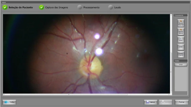
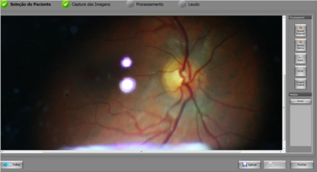
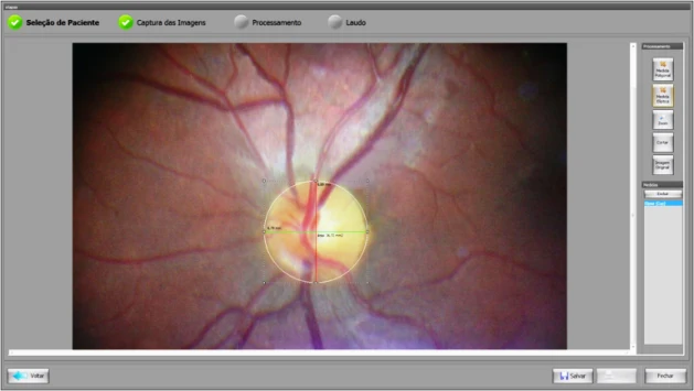
Example images show the left optic nerve of a 32-year-old volunteer and the right optic nerve of a healthy 40-year-old volunteer, both photographed without dilation. The processing screen measured optic-disc and optic-cup areas and their horizontal and vertical diameters, with image tools for detailed optic-nerve analysis.
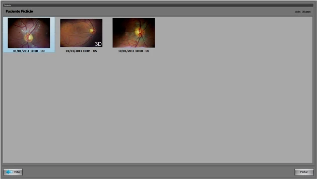
The image database identified stereoscopic pairs with a 3D marker. Saving stereo images as linked pairs supported later review and report reprinting without ambiguity about the paired images.
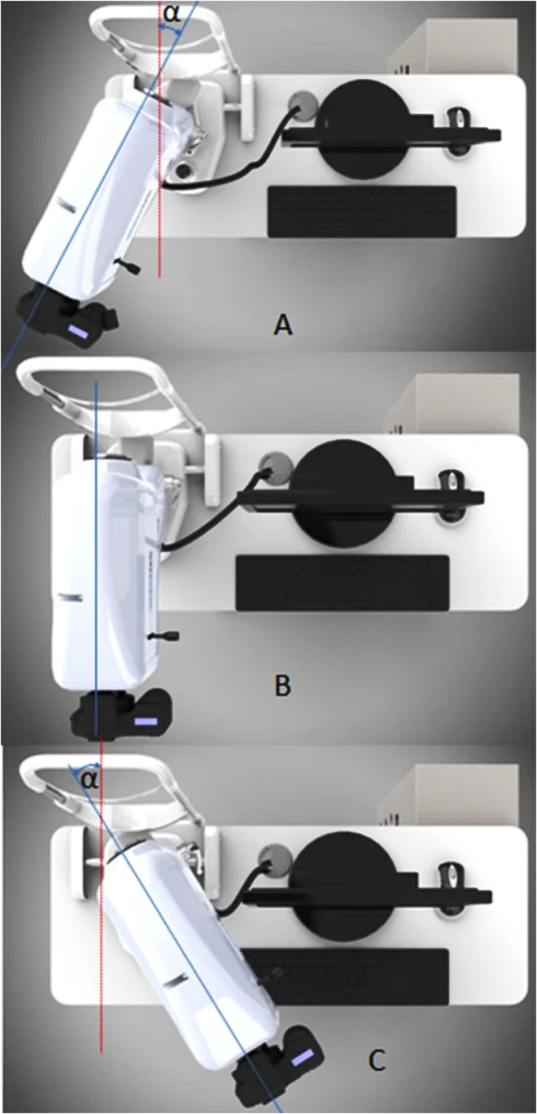
The mechanical design provided vertical tilt and optical-head rotation for stereo acquisition and wider-field imaging. The archived text gives vertical tilt of ±15°, then refers to both ±20° and 60° to each side for rotation in different figure descriptions. These inconsistent historical rotation values require confirmation against the engineering drawings before being treated as a specification.
Quantitative optic-disc parameters included mean disc diameter in millimeters, total disc area and cup area in mm², cup shape, cup volume, cup-to-disc area ratio, horizontal cup-to-disc diameter ratio and vertical cup-to-disc diameter ratio. These measurements appeared in the processing screen, report screen and printed report. The source includes instrument views, software screens and a technical-specification PDF.
Instrument, imaging & software gallery
BROCHURES / VIDEOS / SCIENTIFIC REFERENCES
Explore HOLTZ.
Instrument videos & recorded coverage
Original instrument documents & sources
Brochures and manuals link to English EVOptica editions. Historical recordings, source citations and legacy interface screenshots may retain their original language. External archive links may no longer be available.