THE EVOPTICA INNOVATION ROADMAP
The next generation
starts with a better question.
How can we learn more from light, measure more of the eye and turn complex data into a clearer examination? Our development priorities begin with proven engineering experience and new questions.
CHROMA / DEVELOPMENT DIRECTION
Chromatic insight. A renewed platform.
Building on the founders’ patented chromatic Placido-disc approach, our development direction combines renewed acquisition hardware with updated reconstruction, visualization and examination workflows.
Corneal shape reconstruction, robust pattern interpretation and optical-quality analysis are the engineering priorities. Future performance will be established through verification and validation.
Explore the platform’s foundation ↗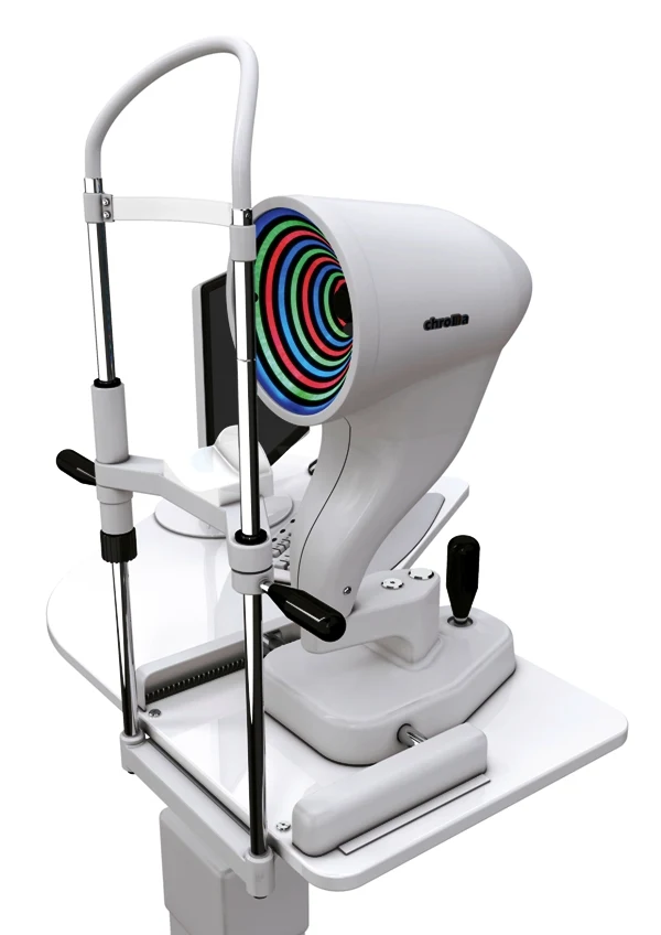 EARLIER PLATFORM / ROADMAP REFERENCE
VON / DEVELOPMENT DIRECTION
A new direction for visual-field stimuli.
We are exploring a chromatic laser-beam stimulus architecture as the next step beyond the earlier LED-based VON perimeter. The development goals are more precise stimulus delivery and improved contrast control.
These are design objectives, not demonstrated clinical performance claims. Optical-radiation safety, calibration, repeatability and clinical evaluation are part of the development work ahead.
Explore the platform’s foundation ↗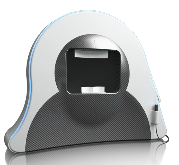 EARLIER PLATFORM / ROADMAP REFERENCE
HERON / DEVELOPMENT DIRECTION
More of the endothelium. More connected analysis.
Our next-generation specular microscopy direction targets improved scanning across more endothelial regions, together with updated acquisition guidance, cell-analysis tools and software workflows.
Building on the earlier multi-position HERON architecture, the goal is a broader view of the endothelial mosaic. Coverage, repeatability and analysis performance will be established during development.
Explore the platform’s foundation ↗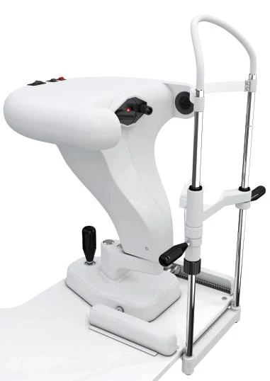 EARLIER PLATFORM / ROADMAP REFERENCE
A BROADER OPTICAL HORIZON
One platform of expertise.
Many paths to discovery.
Our archive extends to ocular aberrometry, Scheimpflug imaging, optic-disc reconstruction, pupillometry, visual acuity, low vision, cross-linking and optical correction research.
Additional work includes OCT biometer–topographer integration and image-analysis research for dry-eye assessment.
From an idea to an evaluated instrument
- Define the clinical question and intended use.
- Model the optical and instrument architecture.
- Build and integrate engineering prototypes.
- Verify performance, safety and software behavior.
- Evaluate the clinical workflow and prepare the production configuration.